Search This Blog

Friday, April 29, 2011

Soft tissue calcification

Anatomical image above shows the menisci, the cartilageneous disc material which acts as shock absorber for the knees.
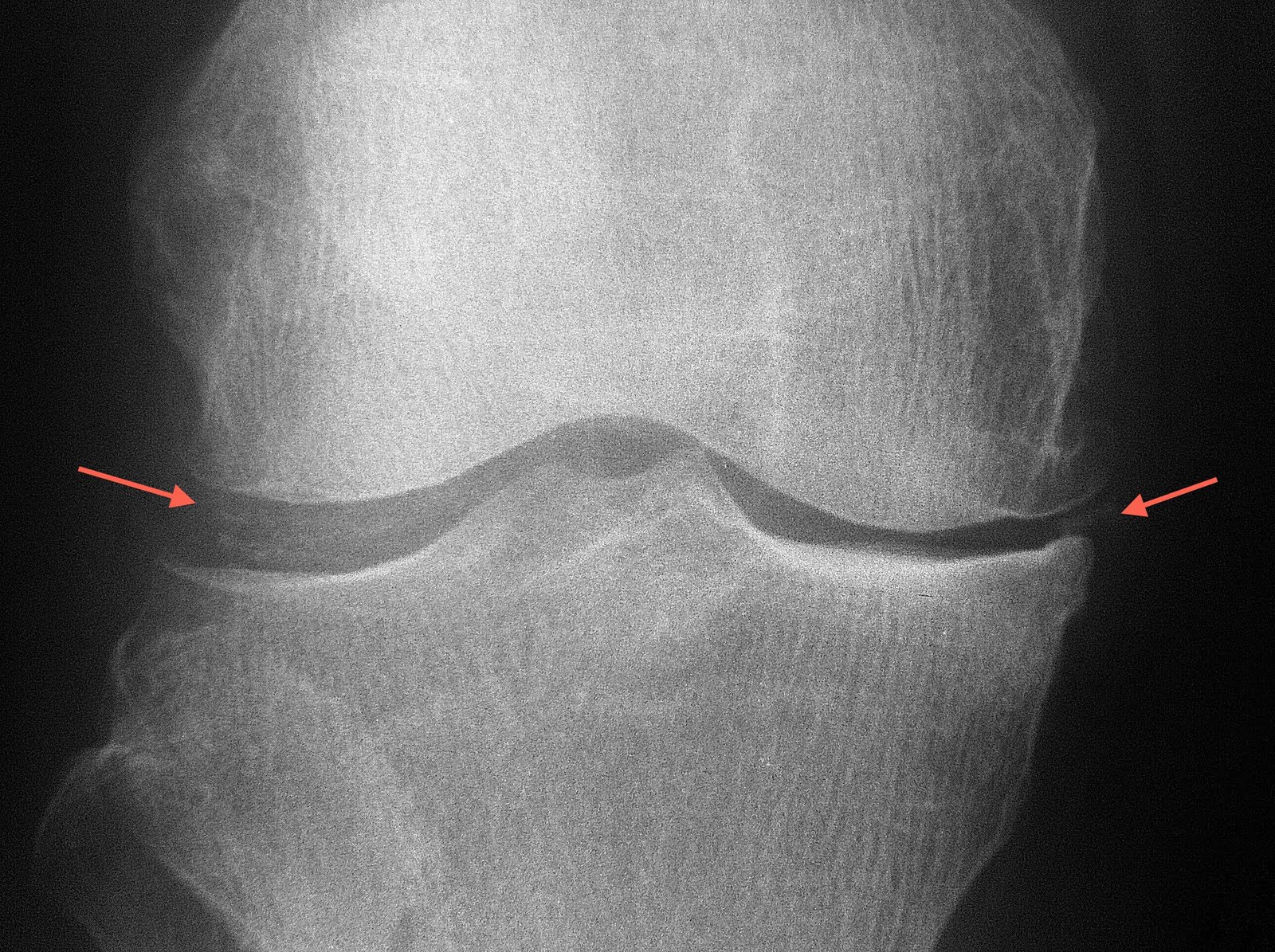
Radiograph of the patient shows calcification of the meniscus. This condition may be observed in patients with gout, although is not specific.

Motion artifact on MRI

MRI may be a very good diagnostic tool, but is subjected to limitations as well. One of the most common problem encountered is movement. Patient in pain often cannot hold still for long period of time, which is unfortunately required during MRI scans. A typical scan with MRI is at least 20 minutes, which to some, is already too long to bear. Image above shows curve signal in the shoulder MRI picture (coronal oblique STIR) due to motion artifact.


Repeated image after advising the patient to hold still saw elimination of such artifact.

Clinicians should provide adequate analgesia to patients prior to MRI scan. Some patients in severe discomfort may benefit from sedation administered prior to the scan.

Incidental finding on CT brain


Midline hyperdense mass on plain CT arising from the interhemispheric falx is in keeping with meningioma. This is the most common benign intracranial tumour. The tumour grows at a very slow pace (like in this patient) which is usually asymptomatic. This patient did the scan to exclude infarct and the meningioma was an incidental finding, despite the size of the tumour.

Big heart

The heart is contained within the pericardial sac. Image above shows the pericardial sac in situ.

Image above shows removal of the pericardial sac revealing the heart.


This young girl with connective tissue disease shows "water bag" sign, which indicates presence of pleural effusion. This sign is due to gravitational accumulation of the pericardial fluid towards the dependent base of the heart, hence appearing as if a bag filled with water is being laid on a flat surface.

Implant assessment


Assessment of orthopaedic implant constitutes part of the reporting "to-do-list". In this radiograph, there is loosening of the proximal cortical screw noted. Other complications to watch out for include fracture of the metallic device or infection.

Avulsion fracture

The posterior cruciate ligament attaches to the posterior aspect of the tibial plateau.

Avulsion fracture at the site of the tibial attachment of the PCL can occur with trauma of variety mechanism. Treatment involves reducing the avulsed fragment into the tibia whilst ensuring that the PCL is not torn.

Another pertinent finding in the radiograph above is the opacification of the infrapatellar (Hoffa) fat pad which indicates presence of knee effusion.

Soft tissue tumor


Radiographs above showed a huge tumour around the elbow joint with calcifications which resemble "rings and arcs", suggestive of chondroid tumour.


MRI of the joint (T1-Gadolinium enhanced) showed an angry looking mass with heterogeneous enhancement. Although MRI is not diagnostic in most soft tissue tumour, it gives important information with regards to the aggressiveness of the lesion (e.g. muscular, neurovascular bundle and bone involvement) as well as the compartment involved in the limbs. The information will prove useful for the surgeon to plan for surgical removal.