Search This Blog

Wednesday, June 8, 2011

Quality matters

During one of my cycling trips, I reached the Laila Taib college building where the old Sibu airport used to be. As the skies were casting a beautiful sunset behind the college building, I stopped and attempted to capture a picture of the setting sun. When I focused at the skies, the building was dark and vice versa. Guess I need a filling light big enough to fill the college building if I were to catch the magnificent sunset as well.

Anyway, this is one example of the importance of the work of radiographers. I've always likened radiographers as photographers and radiologists / doctors, the film critic. Should radiographers mess up with their exposure factors and produce suboptimal images, no eagle eyes of the radiologist can pick up subtle abnormality.

Big heart

Cardiomegaly, or enlarged heart size on chest radiograph, is a non-specific finding. Besides attributing this appearance to just congestive heart failure, there are many other causes as listed below:

1. False positive as in patient with pectus excavatum.
2. Pericardial effusion due to various causes.
3. Cardiomyopathy.
4. Valvular heart disease.
5. Ventricular aneurysm.
6. Mimickers: paraspinal mass or mediastinal mass.

Therefore it is important that adequate history is provided to the radiologist to come to a sensible conclusion.

The picture above shows an obvious enlarged heart which is due to congestive heart failure.

In order estimate the heart size (or cardiothracic ratio/CTR), it is important that the patient take a full breath to fully expand the lungs. Radiographic assessment can be made to assess if the lungs are indeed adequately expanded. A general rule of thumb is that the heart size should not be more than half the width of the greatest transverse diameter of the chest. Nonetheless this is only a rough estimate and should be correlated with the clinical presentation and other tests (eg. echocardiography, ECG etc.).

Some studies have also recommended that CTR of 0.55 to be considered normal for Asia pacific population due to smaller body habitus in this group of people.

Anterior knee pain in a young patient


This young patient presented with pain in the region of tibial tuberosity region. He is active in school and participated in a number of sports activity.

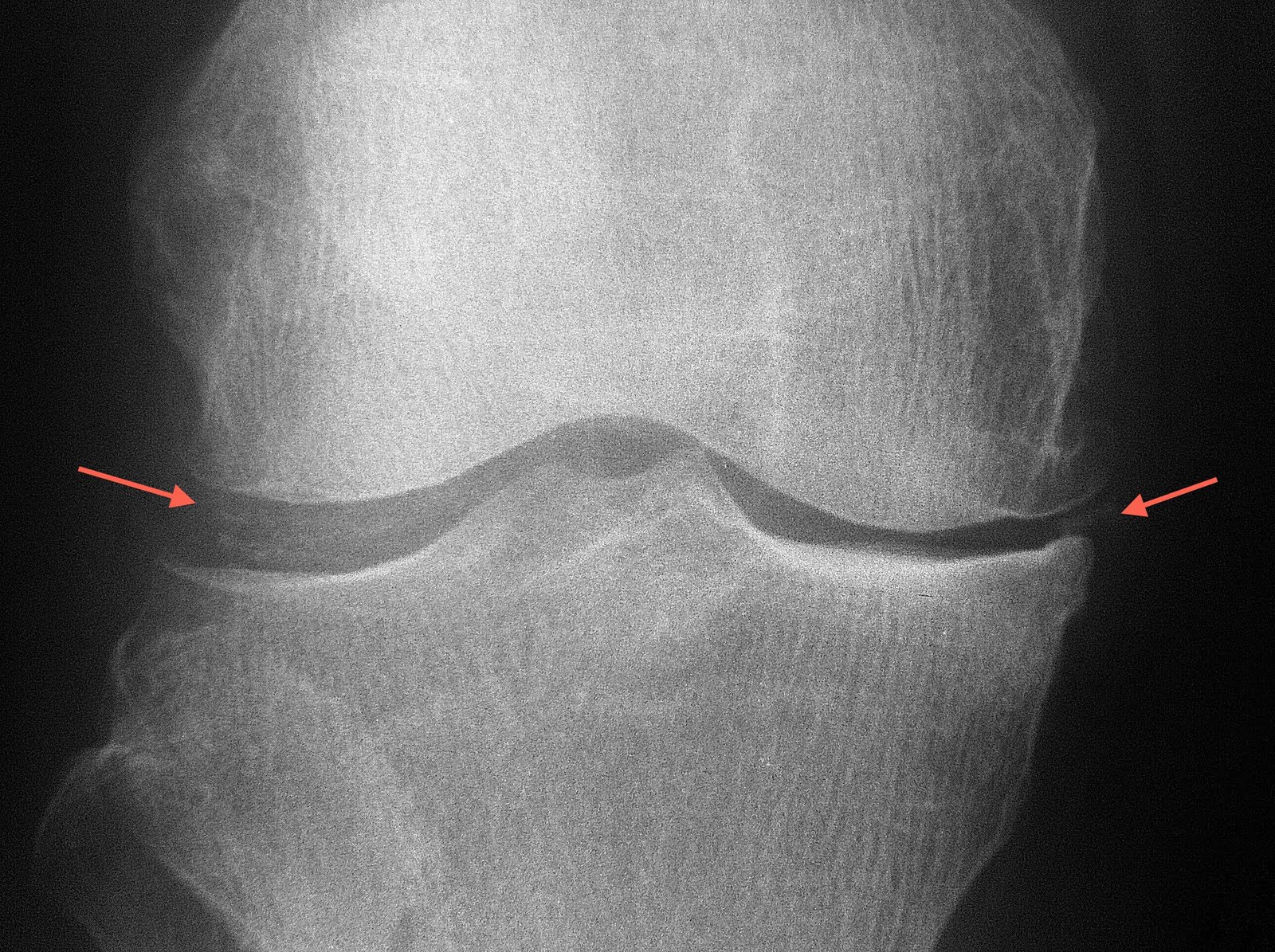
Can you spot the abnormality marked with the arrow?

The lateral knee radiograph shows separation of the ossicles from the ossification centre of the tibial tuberosity. One may also seen soft tissue thickening anterior to the tibial tuberosity.

The diagnosis is Osgood Schlatter disease. It is ne of the most common causes of knee pain in the adolescent. Consisting of pain and edema of the tibial tubercle (and hence this is an extra-articular disease), Osgood-Schlatter disease is generally a benign, self-limited knee condition associated with traction apophysitis of the tibial tubercle due to repetitive strain on the secondary ossification center of the tibial tubercle.

The aetiology of the condition is thought to be due to bone growth which is faster than soft tissue growth, resulting in muscle tendon tightness across the joint and loss of flexibility.

During periods of rapid growth, stress from contraction of the quadriceps is transmitted through the patellar tendon onto a small portion of the partially developed tibial tuberosity. This may result in a partial avulsion fracture through the ossification center. Eventually, secondary heterotopic bone formation occurs in the tendon near its insertion, producing a visible lump. Approximately 25% of patients have bilateral lesions.

Treatment is conservative with immobilization and steroid injection for symptomatic relief.

Possible complications that can follow include nonunion of bone fragment, patellar subluxation, chondromalacia, avulsion of patellar tendon, genu recurvatum.

Friday, April 29, 2011

Soft tissue calcification

Anatomical image above shows the menisci, the cartilageneous disc material which acts as shock absorber for the knees.
Radiograph of the patient shows calcification of the meniscus. This condition may be observed in patients with gout, although is not specific.

Motion artifact on MRI

MRI may be a very good diagnostic tool, but is subjected to limitations as well. One of the most common problem encountered is movement. Patient in pain often cannot hold still for long period of time, which is unfortunately required during MRI scans. A typical scan with MRI is at least 20 minutes, which to some, is already too long to bear. Image above shows curve signal in the shoulder MRI picture (coronal oblique STIR) due to motion artifact.


Repeated image after advising the patient to hold still saw elimination of such artifact.

Clinicians should provide adequate analgesia to patients prior to MRI scan. Some patients in severe discomfort may benefit from sedation administered prior to the scan.

Incidental finding on CT brain


Midline hyperdense mass on plain CT arising from the interhemispheric falx is in keeping with meningioma. This is the most common benign intracranial tumour. The tumour grows at a very slow pace (like in this patient) which is usually asymptomatic. This patient did the scan to exclude infarct and the meningioma was an incidental finding, despite the size of the tumour.

Big heart

The heart is contained within the pericardial sac. Image above shows the pericardial sac in situ.

Image above shows removal of the pericardial sac revealing the heart.


This young girl with connective tissue disease shows "water bag" sign, which indicates presence of pleural effusion. This sign is due to gravitational accumulation of the pericardial fluid towards the dependent base of the heart, hence appearing as if a bag filled with water is being laid on a flat surface.