Search This Blog

Friday, April 29, 2011

Soft tissue calcification

Anatomical image above shows the menisci, the cartilageneous disc material which acts as shock absorber for the knees.
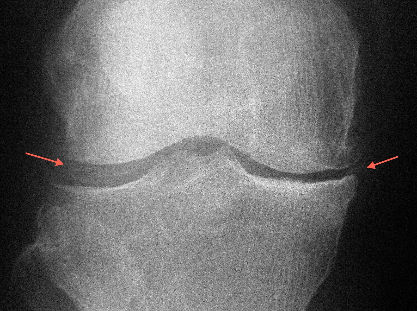
Radiograph of the patient shows calcification of the meniscus. This condition may be observed in patients with gout, although is not specific.

Motion artifact on MRI

MRI may be a very good diagnostic tool, but is subjected to limitations as well. One of the most common problem encountered is movement. Patient in pain often cannot hold still for long period of time, which is unfortunately required during MRI scans. A typical scan with MRI is at least 20 minutes, which to some, is already too long to bear. Image above shows curve signal in the shoulder MRI picture (coronal oblique STIR) due to motion artifact.


Repeated image after advising the patient to hold still saw elimination of such artifact.

Clinicians should provide adequate analgesia to patients prior to MRI scan. Some patients in severe discomfort may benefit from sedation administered prior to the scan.

Incidental finding on CT brain


Midline hyperdense mass on plain CT arising from the interhemispheric falx is in keeping with meningioma. This is the most common benign intracranial tumour. The tumour grows at a very slow pace (like in this patient) which is usually asymptomatic. This patient did the scan to exclude infarct and the meningioma was an incidental finding, despite the size of the tumour.

Big heart

The heart is contained within the pericardial sac. Image above shows the pericardial sac in situ.

Image above shows removal of the pericardial sac revealing the heart.


This young girl with connective tissue disease shows "water bag" sign, which indicates presence of pleural effusion. This sign is due to gravitational accumulation of the pericardial fluid towards the dependent base of the heart, hence appearing as if a bag filled with water is being laid on a flat surface.

Implant assessment


Assessment of orthopaedic implant constitutes part of the reporting "to-do-list". In this radiograph, there is loosening of the proximal cortical screw noted. Other complications to watch out for include fracture of the metallic device or infection.

Avulsion fracture

The posterior cruciate ligament attaches to the posterior aspect of the tibial plateau.

Avulsion fracture at the site of the tibial attachment of the PCL can occur with trauma of variety mechanism. Treatment involves reducing the avulsed fragment into the tibia whilst ensuring that the PCL is not torn.

Another pertinent finding in the radiograph above is the opacification of the infrapatellar (Hoffa) fat pad which indicates presence of knee effusion.

Soft tissue tumor


Radiographs above showed a huge tumour around the elbow joint with calcifications which resemble "rings and arcs", suggestive of chondroid tumour.


MRI of the joint (T1-Gadolinium enhanced) showed an angry looking mass with heterogeneous enhancement. Although MRI is not diagnostic in most soft tissue tumour, it gives important information with regards to the aggressiveness of the lesion (e.g. muscular, neurovascular bundle and bone involvement) as well as the compartment involved in the limbs. The information will prove useful for the surgeon to plan for surgical removal.

Calcific bursitis

Bursae are fluid fill sacs located near the tendons in major joints. Bursitis is a relatively common cause of joint pain associated with movement. When bursitis is chronic, calcium deposit can be noted within the bursa, hence termed calcific bursitis. Shown in picture above is an example of iliopsoas bursa which has calcified.


Shown in images of the knee above, the calcified suprapatellar bursa. Patient with calcific bursitis who presents with recurrence pain maybe candidate for surgical removal of the bursae.

Not all opacities are pathological

Can you spot the vague opacity in the centre of the chest X-ray above.

Picture below outlines the opacity for you.

Whenever an opacity is seen in a chest X-ray, it is important to consider external artifact that can give rise to such appearance. Radiographically, anything external (on the skin) will give a lucent halo around the opacity due to normal air that is trapped between the object and the skin. In this case, the patient was actually wearing pendant made of clothing material.

Hip pain

Femoral head has unique blood supply with a inconsistent supply from the artery of ligamentum teres and the arterial ring from the neck region. Interruption of blood supply brought on by femoral neck fracture can potentially result in avascular necrosis of the femoral head.

Another common cause of AVN includes steroid therapy. However, up to a quarter of patients with AVN have no identifiable causes.


Three plain Xray images showing AVNs in patients who are unaware that they have the condition. These patients present with hip pain, initially thought to be due to degenerative change.

Limitation of plain radiography in bone infection


A young diabetic man presented with chills, rigors and proximal calf pain for 1 week duration. Local palpation revealed local tenderness in the knee area, particularly at the proximal tibia. Plain X-ray (above) shows small area of ill-defined lucent area in the metadiaphyseal region of the proximal tibia. There was no periosteal reaction noted. No obvious soft tissue abnormality.


MRI of the calf shows massive area of marrow oedema involving the proximal tibia as shown in the image above (coronal STIR sequence).

The imaging diagnosis is early osteomyelitis.

This case illustrates the sensitivity of MRI in detecting early bone infection. As with other diagnoses, early detection afford early intervention which limits the resultant damage.

Bronchial abnormality on imaging


Elderly man with productive cough. Chest X-ray shows non-tapering peripheral airway dilatation along with peribronchial thickening.

These findings are consistent with that of bronchiectasis.

Plain radiograph can pick up late disease as per this case. Early bronchiectasis may be elusive of plain X-ray. HRCT thorax is the modality of choice in demonstrating bronchiectasis with greater sensitivity.


Picture above is the HRCT of another patient showing bronchiectasis with additional finding of peripheral honeycombing and subpleural fibrosis due to idiopathic fibrosing alveolitis.

HRCT (high resolution CT scan) of the thorax is a special protocol for examining for airway and interstitial diseases involving the lungs. In this technique, non-overlapping, thin collimation of the primary beam with high resolution algorithm are used. This protocol is different from the typical CT thorax which may not be able to demonstrate interstitial disease as clearly as HRCT.

Cost of running the medical imaging department


I used to have some doubts myself about how medical centres charge patient for imaging studies, which at times may seem like a lot. For e.g. an MRI of the joint typically costs around RM600-RM800.

After having worked in a private healthcare facility for a couple of months, I am beginning to appreciate the cost “behind” operating these mega machines.

To start with, the scanners usually cost about RM1 to 2 million. Renovation of the room is another 100 – 300 thousand ringgit. On top of that the annual service contract is typically about 5-10% of the cost of the machine. Some of the accessories used are non-serviceable (i.e. they are replaced if they are found to be faulty and cannot be fixed).

Featured in this article is the image of an MRI coil which was faulty recently producing images with a dark band on the right side. Unfortunately the coil’s warranty period has lapsed and it is non-serviceable. The construct of the coil looks simple enough with outer casing made of fiberglass material, few stripes of steel films, coils, transistors and cables. Guess how much this coil cost? A whooping RM50,000.00!

Considering all these additional costs, what patient pays may not even be enough to cover the initial and maintenance costs. But such is the nature of running the imaging department. We provide support to the clinicians who can in turn decide the best way to manage the patients.

Friday, April 22, 2011

Shoulder Bursitis

Picture on the left shows the expected location of the subcoracoid bursa in the sagittal view.

MRI of a middle-age lady with painful shoulder and reduction of range of movement suspected of rotator cuff disease. MRI showed fluid within the subcoracoid bursa, which in this case, communicates with the subdeltoid/subacromial bursa. The rotator cuff tendons and muscles are unremarkable. Labrum is intact.

In the absence of ratator cuff disease, finding of fluid in the bursae is suggestive of bursitis.

Communication between these bursae in the shoulder occurs in up to 20% of normal population.

Bursitis can be confidently diagnosed with correlation of clinical and imaging findings. For imaging, MRI is the recommended modality as it demonstrates the expected location of the bursae well and MRI is sensitive to increasing fluid present within these bursae.

Convenience and quality

A middle age man from a town far away came to our department with acute pain to rule out urinary calculus. During routine questioning, we noted that he has just taken his meal mere hours ago. By right, we routinely prep the bowels and fast the patients prior to IVU prior to the study. But in this case, we need to make the exception in view of the logistic reason and the urgency of the case. Shown in the image above is the preliminary film with calculi in the right urinary system. As the preliminary film shows not much of bowel gas, the study was proceeded.


Unfortunately as the study progressed, the bowel gas just kept on increasing to the extent that it almost obscure the view. Fortunately fast bolus injection of pre-warmed contrast was given, hence the concentration of the contrast excreted into the urinary system was sufficient to enable visualization of the urinary tracts beyond the bowel gas.

The lesson learnt in this case is the importance of fasting. Non-fasting patient can have increasing bowel gas which may obscure the opacified urinary tracts. It may not be justifiable to sacrifice quality for convenience. But when it comes to answering important clinical questions, exceptions may need to be made, as per this case.

Fatty Liver Disease

Liver is the largest organ in the human body. It has over 500 vital functions which can be summarized briefly as detoxification, production of various proteins & enzymes and storage functions for sugar & fat.

A growing concern for liver health due to the fast food and sedentary lifestyle era is a condition called hepatic steatosis (fatty liver). In this condition, large vacuoles of triglyceride fat molecules are accumulated within the liver cells (hepatocytes).

Other causes of fatty liver include:
Alcohol
Diabetes mellitus
Obesity
Steroids therapy
Chronic viral hepatitis
Chemotherapy

In some people, fatty liver can progress to inflammation of the liver cells (steatohepatitis) and even resulting in scarring of the liver (cirrhosis) and cancer formation (hepatocellular carcinoma). The long term harmful effects of fatty liver has just be discovered in the last decade through various clinical studies.

Fatty liver can be detected using ultrasound scanning of the abdomen. Normal liver has sharp edges with relative dark appearance (low echogenicity) as shown here in the images with blue arrow. However, patients with fatty liver will demonstrate bright liver (high echogenicity) as shown here in images with red arrows. Occasionally, the liver can be swollen as well (hepatomegaly) giving rise to epigastric discomfort.

Fortunately fatty liver is a reversible condition. The importance lies in early detection of disease and close follow-up with biochemical profile (liver function tests).

Wednesday, April 20, 2011

Breast Imaging: mammogram or ultrasound?


The breast can be investigated using a few methods, which include ultrasound, X-ray (mammography), MRI and thermography. At the moment, breast ultrasound and mammography remain as the main modalities frequently used.

So how does one choose which one to go for?

Women undergoing asymptomatic screening for breast cancer should undergo mammography as the first line. This is because mammography can detect microcalcifications as seen in DCIS (ductal carcinoma in situ), an early manifestation of breast cancer. DCIS can turn into invasive cancer in up to 50% of patients within the period of 10 years.

Ultrasound on the other hand, cannot pick up microcalcifications as good as mammography.

During mammography, another important finding is presence of a mass. If a mass or nodule is detected, it will be shown as a "white" shadow. Mammography can assess the margins of the lesion, the degree of 'whiteness" (density) of the lesion, presence of spicules, architectural distortion.

However, some women have very dense breasts, i.e. very thick glandular tissue. Hence the normal glandular tissue can obscure the mass lesion as both normal glandular tissue and mass lesion appear as white area on mammogram. Therefore patient with dense breasts (Bi-Rads category 3 and above) are routinely offered complimentary ultrasound to exclude mass lesion. Dense breasts do not hide the microcalcifications on mammogram, thus is not a concern for missing DCIS.

Picture on the left is a Peruru Mammography Unit which the centre currently owns and operates. It has won gold prize in Good design award in 2007 due to its ergonomic design and the patient comfort it provides.


Example of a mammogram. Arrow points at a well defined mass lesion which turns out to be a benign neoplasm (fibroadenoma)

Tuesday, April 19, 2011

Hysterosalphingography (HSG)

Netter 2006

HSG remains one of the few techniques which is used to evaluate the patency of fallopian tubes.

It is a simple office procedure done by radiologist using aseptic (sterile) technique which involves insertion of a small sized Foley's catheter into the uterine cavity, followed by introduction of water soluble contrast media into the uterine cavity, thence the patent fallopian tubes and finally into the peritoneal cavity.

The procedure is routinely done during day 4 to day 10 following the cessation of the menses. Patients are required to fast overnight from solid although fluid restriction is not necessary. Patients with documented prior allergy or atopy (e.g. asthma) are routinely premedicated with oral prednisolone with dosage of 40mg 12 hours and 40mg 2 hours prior to the study to avoid contrast adverse reaction. Following the study, pelvic discomfort akin menstrual pain is experienced by some women. Cervical manipulation may induce vasovagal reaction in some, so patients should be accompanied by the partner or some company. If contrast fails to pass through the cornu, IV hyoscine may be administered to induce smooth muscle relaxation. Patients are also advised to bring sanitary pad as spotting due to cervical manipulation is common.

Second picture shown here is an example of a normal HSG where both tubes are normal in caliber and there is spillage of the contrast into the peritoneum indicating patency of the fimbrael ends. Abnormal findings which can be diagnosed via HSG includes blocked tube, hydrosalphinx, abnormality of the uterine cavity (uterine synechiae, endometrial polyp or fibroid) or congenital abnormality of the uterus. All these abnormalities can present as risk factors for infertility.

HSG is provided by this centre via strict appointment basis only.

Bruised bone?

Yes the bones can be bruised! The medical term for this condition is trabecular microfracture. Before the advent of MRI, patients presenting with persistent pain after trauma without demonstrable fracture are assumed to have occult fracture. With special scan sequence on MRI (water suppressed T2WI), bone bruise can be easily depicted, as shown by the image on the left.

To qualify a lesion as trabecular microfracture, no cortical fracture must be demonstrable.

In the old times, to demonstrate this, patients have to be subjected to nuclear medicine scan which involves radiation and injection of radiopharmaceutical agent. Nowadays with MRI, the diagnosis can be obtained within minutes without any injection or radiation exposure.

Trabecular microfracture is often self limiting, with some delay in MR resolution compared to clinical improvement of symptom, which is typically within two to three months. Subchondral bone bruise (as index case) increases the risk of articular cartilage injury as the lesion is usually due to compaction of the bones. Internal knee derangement is also possible with such finding (e.g. meniscal or cruciate ligament tear) and should be excluded as well.

Imaging the articular cartilage

Imaging the articular cartilage has not been an easy task. Being radiolucent, it cannot be seen with conventional radiography or CT scan. It is inaccessible to ultrasound.

Often the evidence of worn out cartilage is gathered from plain radiograph when the joint space is reduced. But this is already a late presentation of disease.

MRI proves to be an invaluable tool for assessing articular cartilage, particularly in the setting of trauma. On GRE Water Image as shown in the images in this article, normal articular cartilage shows as a high signal (bright) rim that surrounds the articular surfaces. On MRI, thinning, fissuring or focal defect can readily be demonstrable.


This image shows full thickness cartilage loss (arrowheads) here in the medial condyle in a patient with recent road traffic accident.

Identifying early cartilage loss is important to help patients take active measures to reduce further damage to the cartilage. Interventions may include lifestyle changes like weight modification or use of supportive devices, pharmacological approach or surgery.

In this centre, our MRI is fully capable of performing GRE Water image to evaluate the articular cartilage thickness. Most of the times, the scan done is in conjunction with assessment of the other internal knee structures such as the menisci and ligaments.