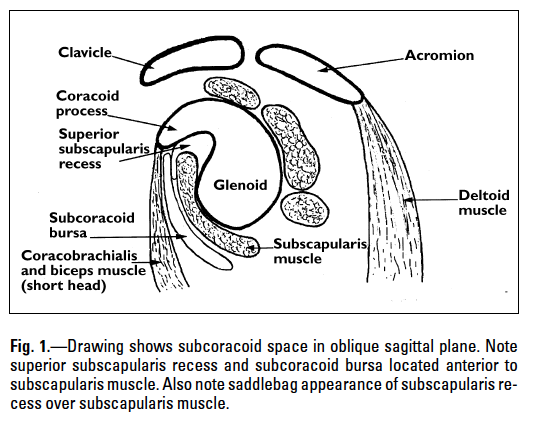
 Picture on the left shows the expected location of the subcoracoid bursa in the sagittal view.
 MRI of a middle-age lady with painful shoulder and reduction of range of movement suspected of rotator cuff disease. MRI showed fluid within the subcoracoid bursa, which in this case, communicates with the subdeltoid/subacromial bursa. The rotator cuff tendons and muscles are unremarkable. Labrum is intact.
In the absence of ratator cuff disease, finding of fluid in the bursae is suggestive of bursitis.
Communication between these bursae in the shoulder occurs in up to 20% of normal population.
Bursitis can be confidently diagnosed with correlation of clinical and imaging findings. For imaging, MRI is the recommended modality as it demonstrates the expected location of the bursae well and MRI is sensitive to increasing fluid present within these bursae.
No comments:
Post a Comment